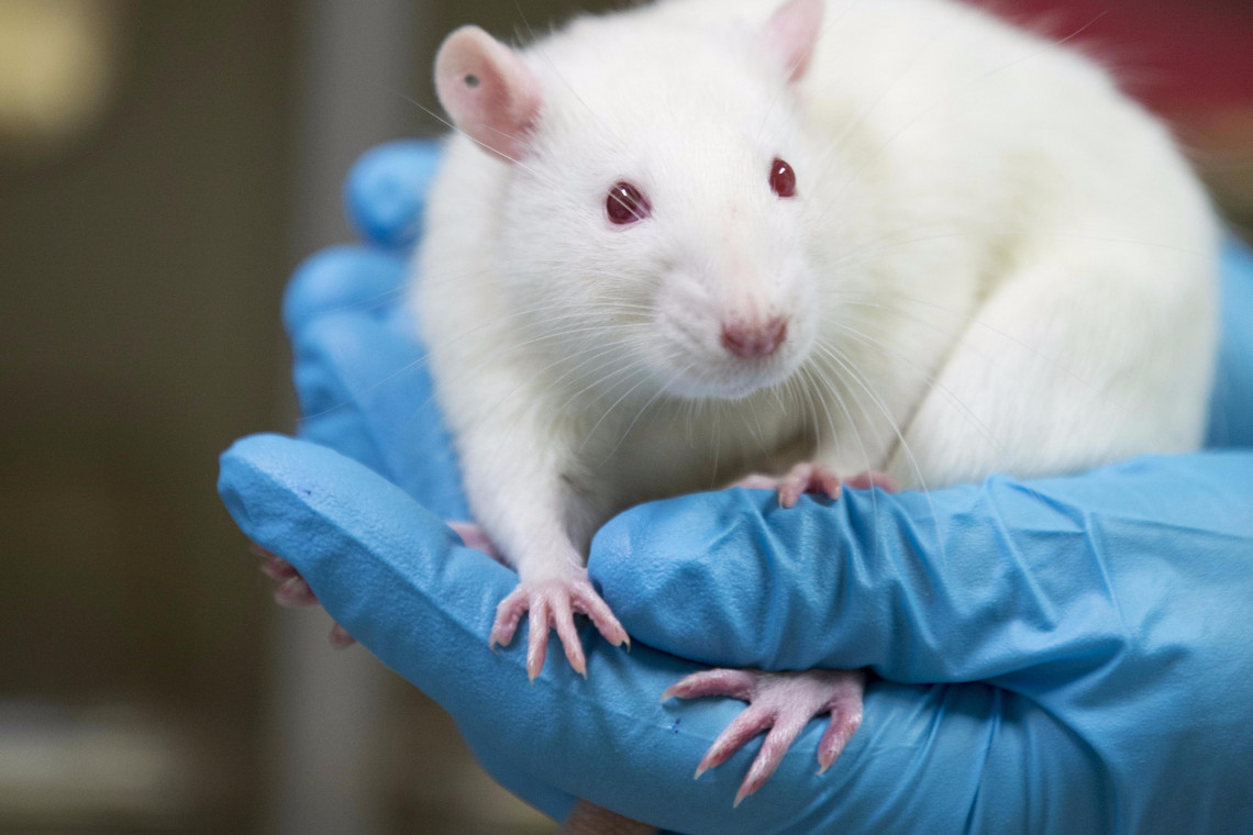
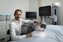
Chris de Korte is professor at the UT and head of the Medical UltraSound Imaging Center at the Radboudumc in Nijmegen. Overall, his work is dedicated to developing new ultrasound techniques and using them to improve diagnosis of cancer and cardiovascular diseases. His most recent research project, in collaboration with UT professor Michel Versluis, is focused on painless and quick detection of breast cancer. This research project has been supported by the UT community, recently attracting 10.000 euros in donations from the Twente University Fund.
Ultrasound and contrast agents
‘Normally the process of detecting breast cancer is quite cumbersome,’ says De Korte. ‘If a woman finds a lump in her breast, mammography and ultrasound scans are performed. If the radiologist suspects that it is malignant tissue, a biopsy is taken. However, in case the lesion remains occult in the ultrasound, an MRI scan is necessary to identify the lesion and take the biopsy. That can mean a week or more until the result is known.’
‘Normally the process of detecting breast cancer is quite cumbersome’
‘We’d like to use ultrasound for all these steps, so that MRI is not necessary and you get the full diagnosis in one day,’ explains the scientist. ‘The key to making this a reality is combining ultrafast ultrasound with elastography and contrast imaging. With elastography we ‘feel’ with ultrasound where hard lesions are located.’
3D scanner
‘We have already developed a new 3D imaging system that gently deforms the breast tissue to detect lesions,’ continues De Korte. ‘We have tested this method in about eighty patients and the results are very promising. We are able to distinguish between benign and malignant lesions. Recently, we developed a cone shaped scanner that rotates around the breast and can scan the full breast in less than five seconds. Now we’d also like to add contrast imaging to even better detect malignant lesions. For that, we like to investigate how the contrast agent exactly interacts with the tumors and how to detect that contrast agent super-sensitively with ultrasound.’

A schematic drawing of the cone scanner and a 3D ultrasound breast scan as an initial result
Finding the right contrast agent for this specific application is, in fact, the next step of the research project supported by the Twente University Fund donations. ‘We really appreciate this extra funding,’ says the professor. ‘The whole project amounts to about one million euros in total, but this is really precious money, money given by individual people, and we want to use it in such a way it has the maximum impact.’
Faster and cheaper
The UT scientist believes that the technique could be used in the clinic within years. ‘If we have sufficient funding, we can really demonstrate how well the method works in five years. After that it is up to the companies to pick it up and incorporate it. However, we already closely collaborate with manufacturers such as Siemens.’
'The method is not only faster, but also a lot cheaper than MRI'
Ultimately Chris de Korte wants to create a method that is better for the women and the healthcare. ‘Instead of waiting for a week for MRI, a woman will know her full diagnosis the same day. You can also use the 3D ultrasound to take a biopsy because you can see in real time where the biopsy needs to be taken. This means much less discomfort and insecurity for women. The method is not only faster, but also a lot cheaper than MRI, which is of course better for the healthcare system. On top of that, with our new technique, more women can be examined, for example also younger women: tumors in this group are often undetectable with X-rays. My dream is to develop an comfortable and accurate breast cancer detection system to improve survival of breast cancer.’